 |
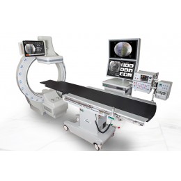
X-RAY systems Biotok XR 3D
3D x-ray imaging in real time.
Fluoroscopic 3D mapping and navigation.
Part number:
Description
Biotok XR 3D is the best choice for diagnostic and interventional cardiology, angiology and other procedures performed under x-ray control. Stereo x-ray generator power up to 15kW provides stereoscopic images of dynamic objects (tools, vessels, heart etc) in real time with excellent quality. A three-dimensional grayscale x-ray image allows to determine the direction of movement of the tool in the body of the patient, more precisely to position, promote and measure objects in a plane and in depth. System naturally discriminates the noise, thereby increasing the information of images. New flat panel detector sizes up to 30 cm, frame rate up to 60 fps and the highest resolution 4K×3K (pixel size of 75 µm) is informative, precise detail without distortion and quality loss, as well as comfortable work in any field of interest.
Biotok XR 3D is not only volumetric visualization in real time, but it is also the fluoroscopic 3D mapping and navigation system.
3D reconstruction in Biotok XR 3D
HIGHLIGHTS
- Increase the informative value and volume of diagnostic information, by obtaining three-dimensional stereoscopic grayscale x-ray images in real-time including moving objects (for example: heart).
- 3D reonstruction, for example heart chambers, by analogy with nonfluoroscopy navigation only by obtaining three-dimensional coordinates of points of the catheters and the surface of the building group in the amount of memorized points.
- Significant simplification and acceleration of work of the surgeon: when navigating and advancing the tool in the x-ray image with the construction of stereo vascular map to advance catheters; at diagnosis (advancement of contrast agent through the vessels) in the volume and dynamics; when detecting low-contrast and geometrically small structures, etc.; accurate positioning and placement of implants.
- Moving and navigation tool in a hazardous environment, to avoid surgical injury and accelerate the rehabilitation period of the patient.
- Definition of Intraoperative vascular perfusion of the contrasting results of the vascular.
- Integration of previously acquired three-dimensional objects or surfaces obtained in the stereoscopic image.
- Pulse X-ray generator and tube with rotating anode power up to 15 kW provide the dynamic STEREO superior-quality images and low radiation dose to the patient.
- Isocentrically motorized-arc provides stable rotation in space with compensation of the inertia in the range up to 240 degrees for 5-20 seconds with a return to the memorized position.
- Touch control.
- Packages "Visualization of vessels"; "Cone-beam tomography" (3D reconstruction); "Synchronization with ECG"; "the Combination of 3D imaging with fluoroscopy, real-time".
- increase the informative value and volume of diagnostic information, by obtaining three-dimensional stereoscopic grayscale x-ray image in real-time including mobile objects (for example: heart).
REAL-TIME ADVANCED IMAGE PROCESSING
Multilevel advanced image processing in real time using the latest developments in the theory of digital filtering and analysisNEW FEATURES IN APPLICATIONS
Angiography;
Endovascular, cardiovascular surgery, radiofrequency ablation, the installation of the occluders;
Implantation;
Scintigraphy;
Definition of perfusion;
Catheterization;
Traumatology, orthopedics, General surgery: installation of implants of joints; fixation of implants in the patient's body; pointing of the instrument; installation of the spokes; pelvis; locking nail; hip pain, hip replacement; knee pain, knee replacement; back pain; and so on…
Biopsy;
Treat and manage pain
Neurosurgery - promotion and navigation tool in traumatic area;
Urology: – positioning and navigation tool in the genitourinary system; improving the detection of deposits in urolithiasis; percutaneous nephrolithotomy; cholangiography;
Bypass surgery;
Neurology;
Head/Spine: neuromodulation; hypophysectomy; spine fixation;
Cone-beam tomography with high quality.