.png) |
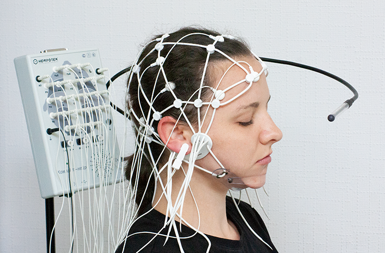
Encephalograph Compact-Neuro 21 channels
21-channel Compact-Neuro encephalograph is intended to perform standard electroencephalography in stationary conditions.
This is an extended version of the electroencephalograph with the increased number of leads. It can be used to perform deep EEG examination in neurological and psychiatric diagnostic, as well as in epileptology. It allows receiving information from the increased number of brain zones in order to detect local changes.
Part number:
Supplier:
NeurotechDescription
An electroencephalograph is a device required to perform the routine (using the most common methods) monitoring of the bio-electric potentials of the human brain following randomly selected scanning protocols. The main purpose of the system os to diagnose epileptical changes in EEG, monitor EEG at night, as well as the dynamic evaluation of the functional activity of the brain. The monitoring can be carried out using any number (up to 21) standard lead electrodes in both monopolar and bipolar modes with simultaneous recording of the patient's state using a web-camera.
The device supports the following features during the real-time usage:
changing scales of time, amplitude, and filter parameters
custom range filters allowing selecting specific frequency ranges
checking the quality of electrodes positioning (impedance measuring)
filtering power line disturbances
choosing bipolar schemes
recording with/without the protocol, custom order of samples
notepad with timestamps and events
The device supports the following features during the real-time usage and after the registration:
spectral analysis
the cartographical analysis in 2D and 3D
correlation analysis
automatic artifact detection
The device supports the following features after the registration of EEG:
automatic searching of ranges with pathologic activity
automatic report generation in text or in table formats
recording results and video to CD
import/export of examination results
EEG signals export in EDF and CSV
remote impression writing using the cloud storage or a network folder
SOFTWARE FEATURES
The convenient database allows fast patient information adding.
Supports parallel recording from two or more cameras.
EEG registration can be carried out with available of with custom protocols.
Quality of the electrode attachment control by measuring the impedance between the electrodes in real-time.
Both reports and examination information can be stored and transferred using the cloud storage.
Automated impression creation for functional batches in the report.
Ability to use digital filters as in real time, as well as after the signal registration.
Results of the examination can be exported in CSV, EDF, and EDF+formats, and processed using the most popular international software: LORETA, EEGLAB, BESA, CURRY etc.
Supports restoration of the EEG data from a flash card in case if the patient went out of the receiver coverage.
Main types of EEG processing:
automatic mode for artifacts identification
automatic searching of ranges with pathological activity, including continuous records
spectral and correlation analysis
the cartographical analysis in 2D and 3D
calculation of the hemispheric asymmetry, index, and the rhythm frequency
TECHNICAL CHARACTERISTICS
Number of EEG channels 21
Computer connection USB
Amplifier working frequency 0-80 Hz
Sampling rate for each channel 512 Hz
RMS-noise (at the input) less than 1,5 mkV
Measured signal amplitude range +/-0,8V
AD converter capacity 24
Electrostatic discharge resistance 8 kW
Power supply +5V (USB port)
Phonostimulation
Impulse volume range 0-2 W
Impulse duration 10 µs — 250 µs
Impulse frequency 0,5 Hz - 60 Hz
Photostimulation
Stimulator type LED
Impulse duration 10 µs — 250 µs
Impulse frequency 0,5 Hz - 60 Hz
SPECIFICATIONS
1. Hardware block (1 pc.)
2. USB cable (1 pc.)
3. Lead, main and spare electrodes (cup) (25 pcs.)
4. Reference electrod in the form of an ear pin (3 pcs.)
5. Main electrode with the band (1 pc.)
6. Universal (adjustable) headset to locate the lead electrodes (2 pcs.)
7. LED lamp for the photostimulation (1 pc.)
8. Web-camera (1 pc.)
9. Electrode gel (1 pc.)
10. Special stand for the device (1 pc.)
11. USB-flash with the system software (1 pc.)
12. Operating instructions (1 set)