 |
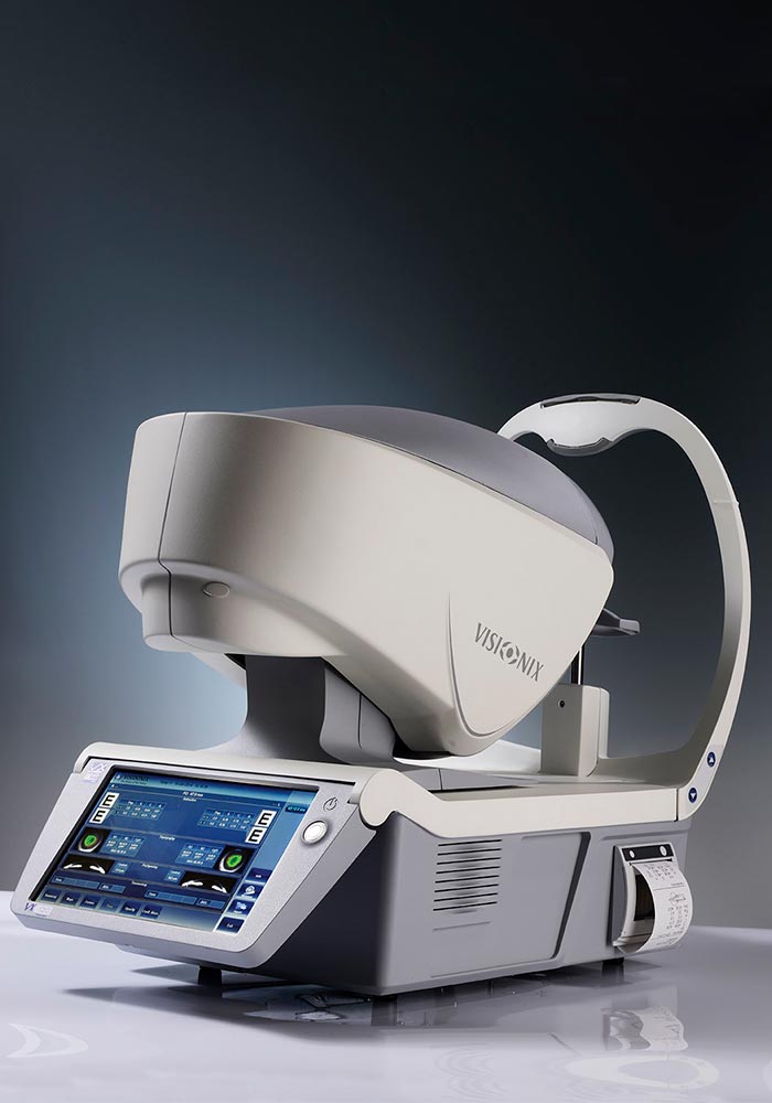
Autorefractometer VX120
The VX 120 is a unique, complete, and fully automatic diagnostic screening device. The VX 120 features variations of refraction, screening for glaucoma, cataracts, corneal pathologies such as keratoconus, and fitting of contact lenses with integrated topography.
Part number:
Supplier:
Visionix RusDescription
The combination of technologies found in the VX 120 are unique (aberrometry, tonometry, topography, Scheimpflug camera, etc.) With full integration in mind, the VX 120 is designed to be able to export measurements and findings and archive your data using WiFi, USB key, office networks, etc.
Fully automated
Fully automatic 3D and R/L eye alignments
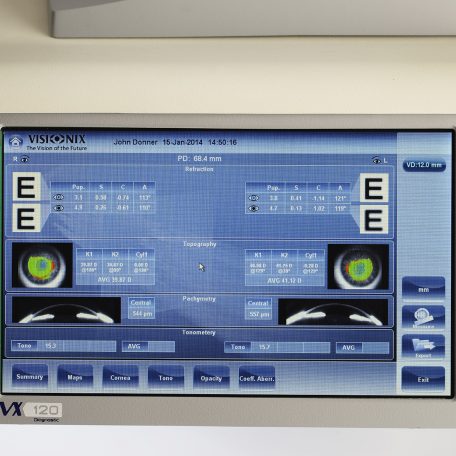
7 types of automatic simultaneous measurements
Operator independent measurements
High reproducibility of measurements
Automatic alignment and measurement which allows
High reliability for measurements
Significant time savings
Optimal comfort based on ergonomic design
Additional customers benefits
Quick detection of refraction, higher order aberrations, and warning indications for measurements outside of normal parameters
Easily transfer patient measurements to the doctor for exam
A refined and highly accurate refraction due to advanced technology and added features
Delegation of tasks
As part of examinations of refraction and detection of high-order aberrations,
possible suspicion of pathologies
General
Alignment……………………….XYZ automatic
Display……………………………10,1” (1 024 x 600) TFT screen
Multi-touch screen
Observation area……………..o 14 mm
Printer…………………………….Integrated black and white – external color available
Medical directive…………….CE MDD 93/42/CE modified
by directive 2007/47/CE
Output…………………………….RS232 / USB / VGA / LAN
AR & power mapping (Wavefront)
Spherical power range……..-20D to +20D
Cylinder power range……….0D to + 8D
Axis………………………………..0 to 180°
Measuring area……………….Min. o 2 mm – Max. 7 mm (3 areas)
Number of measuring points.1,500 points analysis points for pupil of 7 mm
Acquisition time………………0.2 sec
Method…………………………..Shack-Hartmann
Height 540 mm (21.25 in)
Width 320 mm (12.59 in)
Depth 555 mm (21.8 in)
Weight 25 kg (55.1 lbs)
Voltage 100- 240 V AC, 50/60 Hz, 300 W
Pachymetry, IC angle and pupillometry
Method • Scheimpflug
Pachymetry range…………………………………..150-1300 μm
Pachymetry resolution……………………………. +/- 10 microns
IC angle range……………………………………….0°-60°
IC resolution………………………………………….0.1°
Pupil illumination…………………………………..Blue light 455 nm
Retro illumination
Corneal topography
Number of rings…………………………………….24
Number of measuring points…………………..6,144
Number of points analyzed…………………….. More than 100.000
Diameter of covered corneal area at 43D…From 0.75 mm to
more than 10 mm
Diopters measured field………………………….From 37,5 to 56 D
Method…………………………………………………Placido rings
TONOMETER
Measurement range……………………………….7 mmHg to 44 mmHg
Fully automated
Fully automatic 3D and R/L eye alignments
7 types of automatic simultaneous measurements
Operator independent measurements
High reproducibility of measurements
Automatic alignment and measurement which allows
High reliability for measurements
Significant time savings
Optimal comfort based on ergonomic design
Additional customers benefits
Quick detection of refraction, higher order aberrations, and warning indications for measurements outside of normal parameters
Easily transfer patient measurements to the doctor for exam
A refined and highly accurate refraction due to advanced technology and added features
Delegation of tasks
As part of examinations of refraction and detection of high-order aberrations,
possible suspicion of pathologies
General
Alignment……………………….XYZ automatic
Display……………………………10,1” (1 024 x 600) TFT screen
Multi-touch screen
Observation area……………..o 14 mm
Printer…………………………….Integrated black and white – external color available
Medical directive…………….CE MDD 93/42/CE modified
by directive 2007/47/CE
Output…………………………….RS232 / USB / VGA / LAN
AR & power mapping (Wavefront)
Spherical power range……..-20D to +20D
Cylinder power range……….0D to + 8D
Axis………………………………..0 to 180°
Measuring area……………….Min. o 2 mm – Max. 7 mm (3 areas)
Number of measuring points.1,500 points analysis points for pupil of 7 mm
Acquisition time………………0.2 sec
Method…………………………..Shack-Hartmann
Height 540 mm (21.25 in)
Width 320 mm (12.59 in)
Depth 555 mm (21.8 in)
Weight 25 kg (55.1 lbs)
Voltage 100- 240 V AC, 50/60 Hz, 300 W
Pachymetry, IC angle and pupillometry
Method • Scheimpflug
Pachymetry range…………………………………..150-1300 μm
Pachymetry resolution……………………………. +/- 10 microns
IC angle range……………………………………….0°-60°
IC resolution………………………………………….0.1°
Pupil illumination…………………………………..Blue light 455 nm
Retro illumination
Corneal topography
Number of rings…………………………………….24
Number of measuring points…………………..6,144
Number of points analyzed…………………….. More than 100.000
Diameter of covered corneal area at 43D…From 0.75 mm to
more than 10 mm
Diopters measured field………………………….From 37,5 to 56 D
Method…………………………………………………Placido rings
TONOMETER
Measurement range……………………………….7 mmHg to 44 mmHg